Common Mistakes in Doppler Velocity Mapping and How to Avoid Them
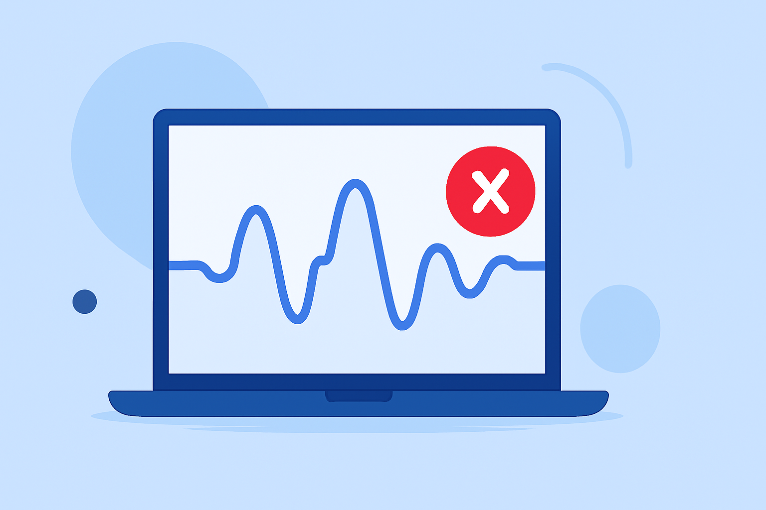
Doppler velocity mapping is a crucial technique in various fields, including cardiology, vascular studies, and astrophysics, for assessing flow velocities and detecting anomalies. However, several common mistakes can compromise the accuracy of these measurements. Understanding and mitigating these errors is essential for obtaining reliable data.
1. Misalignment of the Doppler Beam
Aligning the Doppler beam parallel to the direction of flow is vital. An angle between the beam and flow direction leads to underestimation of velocity. To minimize this error, position the transducer laterally on the patient's chest and adjust the patient's posture to optimize the angle. Avoid using angle correction, as it can introduce additional inaccuracies. (cardioserv.net)
2. Inclusion of Artifacts in Measurements
Artifacts, such as random noise or spurious signals, can distort Doppler tracings. Including these artifacts in measurements can lead to overestimation of velocities. To prevent this, lower the Doppler gain until artifacts disappear while maintaining a clear spectral envelope. Reduce the Doppler scale to improve waveform resolution and avoid tracing fine linear signals beyond the dense spectral display. (cardioserv.net)
3. Confusing Aortic Valve and Mitral Regurgitation Velocities
Mistaking mitral regurgitation (MR) velocity for aortic valve (AV) velocity can lead to significant errors. MR velocity appears earlier in systole and extends through both isovolumic contraction and relaxation phases, while AV velocity begins after isovolumic contraction and ends before isovolumic relaxation. Carefully correlate the timing of the Doppler envelope with the electrocardiogram (ECG) and valve click markers to distinguish between the two. (cardioserv.net)
4. Measuring Post-Extrasystolic Beats
Premature ventricular contractions (PVCs) and the subsequent post-extrasystolic beats can artificially elevate velocities due to increased ventricular filling and contractility. To avoid this, allow the Doppler signal to run for several seconds to capture multiple beats for comparison, and exclude PVC and post-extrasystolic beats from measurements. (cardioserv.net)
5. Aliasing Artifacts
Aliasing occurs when the velocity range exceeds the scale available to display it, leading to reversed flow in high-velocity areas. To correct aliasing, increase the Doppler angle to reduce the measured Doppler shift, or switch to continuous-wave Doppler, which doesn't have a pulse repetition frequency and thus no aliasing. However, continuous-wave Doppler loses depth information, so it should be used judiciously. (epos.myesr.org)
6. Inaccurate Caliper Placement
Incorrect placement of calipers during measurement can lead to overestimation of Doppler velocities. To avoid this, trace the peak velocity along the outer edge of the most intense portion of the signal, excluding fringes and faint linear artifacts caused by the transit-time effect. Ensure that the caliper is positioned at the chin, not at the beard, to accurately measure the modal velocity. (academic.oup.com)
7. Misinterpretation of Doppler Artifacts
Artifacts such as random noise, aliasing, and mirror-image artifacts can lead to misinterpretation of Doppler velocity maps. Understanding these artifacts and their causes is essential for accurate interpretation. Regular calibration of equipment and awareness of potential artifacts can help mitigate these issues. (musculoskeletalkey.com)
Conclusion
Accurate Doppler velocity mapping is essential for diagnosing and monitoring various conditions. By being aware of common mistakes and implementing strategies to avoid them, clinicians and researchers can enhance the reliability of their measurements. Regular training, equipment calibration, and careful technique are key to achieving precise Doppler velocity assessments.
Highlights:
- Continuous-wave Doppler interrogation in valvular heart disease: pearls and pitfalls | European Heart Journal - Imaging Methods and Practice | Oxford Academic, Published on Wednesday, May 20
- Pitfalls in Carotid Doppler Interpretation and How to Avoid Them - Gunabushanam - 2023 - Journal of Ultrasound in Medicine - Wiley Online Library, Published on Wednesday, March 08
- Errors in the estimation of wall shear stress by maximum Doppler velocity - Atherosclerosis, Published on Sunday, February 10